


HQ / Factory : 45, Secheon-ro 7-gil Dasa-eup Dalseong-gun Daegu TEL : +82-1544-2285
MEGAGEN TOWER (International Sales Department) : 609, Nonhyeon-ro, Gangnam-gu, Seoul, Korea TEL : +82-2-6003-2011
FOR CUSTOMER SERVICES
COPYRIGHT(C)MEGA’GEN IMPLANT CO.,LTD.ALL RIGHTS RESERVED
Reduction Malarplasty Using Customized Surgical Stent Based on 3D Virtual Surgery, CAD/CAM, and 3D Printing Technology: Case Series
English 2022-08-25 PDF Lecture 443
Author
마케팅
Date
August 25, 2022
Abstract: The zygomatic bone is a structure that protrudes
symmetrically on both sides of the midface and plays an important
role in the overall aesthetic appearance of the face.
Unlike Caucasians, the mesocephalic facial shape is predominant
in Asians, and therefore, many people have a relatively
laterally developed zygomatic bone. In Asians, when the zygomatic
bone is excessively developed, it gives a strong and
stubborn image, and aesthetically, many people want to reduce
the zygomatic bone because they prefer an oval and slim face.
To reduce the excessive zygomatic bone, a reduction malarplasty
through an intraoral and preauricular approach has been
performed. Although reducing the zygomatic bone is not a big
problem in most cases of symmetric reduction malarplasty, it is
not easy to produce surgical results as intended by the surgeon
in asymmetric malar patients or patients requiring a three-dimensional
(3D) change of zygoma. In addition, because of the
mobility of the zygoma segment, it may be difficult to drill holes
and fix plate after osteotomy. Moreover, these factors can
increase the possibility of malunion or nonunion.
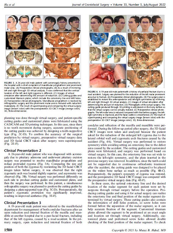
In this study, cutting guides made with the aid of 3D virtual
surgery, 3D printing, and customized titanium plates manufactured
with the computer-aided design/computer-aided manufacturing
technology are used for 8 patients to maximize the
recovery of 3D symmetry and minimize complications through
accurate fixation after surgery. During the surgical procedures,
screw hole drilling and osteotomy were performed using a cutting
guide, and then, the malar segment was fixed by matching
the premade customized plates with the predrilled holes. As a
result of checking the accuracy of the surgery by superimposing
the postoperative 3D cone beam computed tomography image
and virtual surgery data based on the skull base, the 2 images
almost overlapped and no significant differences were observed,
so it was confirmed that the operation was performed exactly as
planned.
When using the 3D technology, it is possible to perform a more
accurate surgery in patients with asymmetry due to congenital
anomalies or trauma as well as simple asymmetry, so it can be
concluded that using the 3D technology can overcome the
limitations and disadvantages of the conventional method as in
the cases in this study. The accurate prediction of soft tissue is
still insufficient, and further research is needed to overcome this
limitation
Key Words: 3D virtual surgery, CAD/CAM, reduction malarplasty,
surgical accuracy